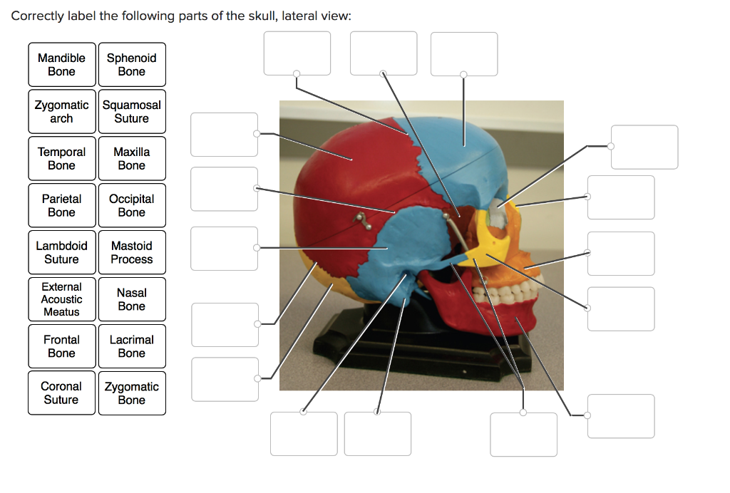
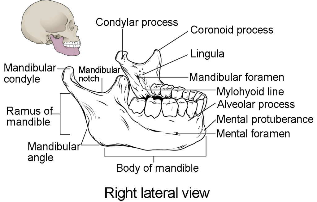
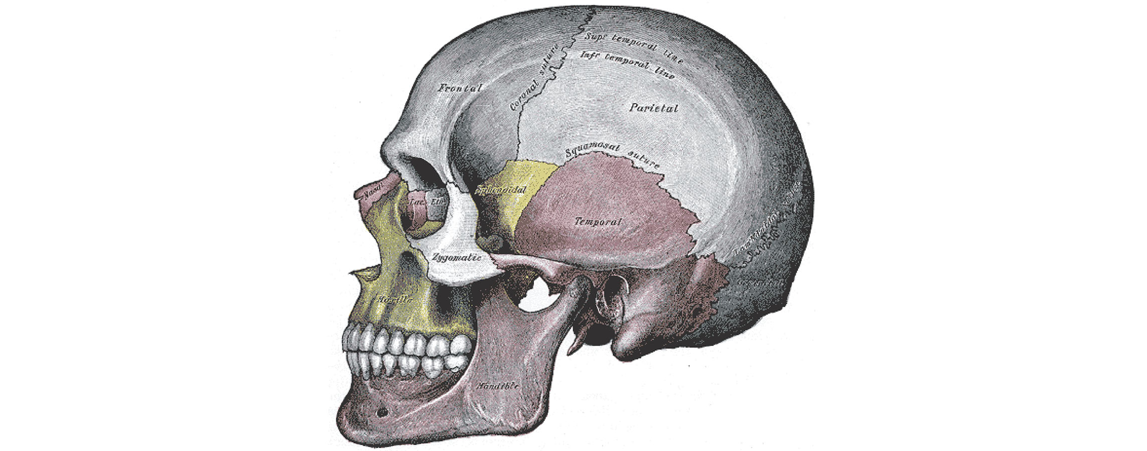
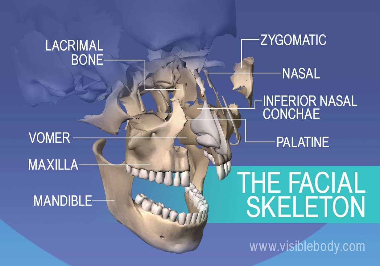
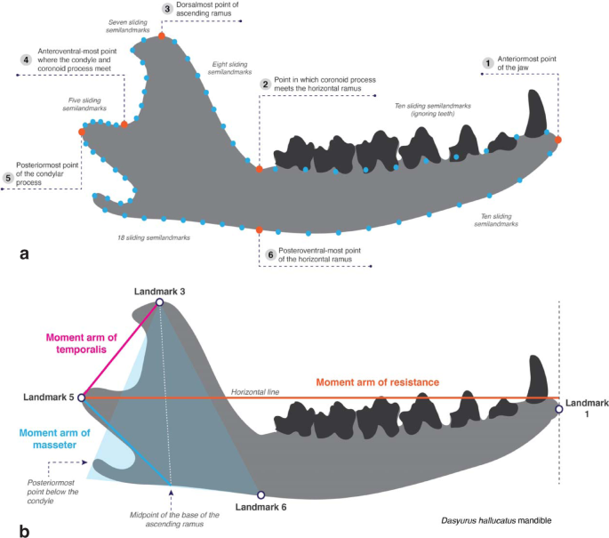
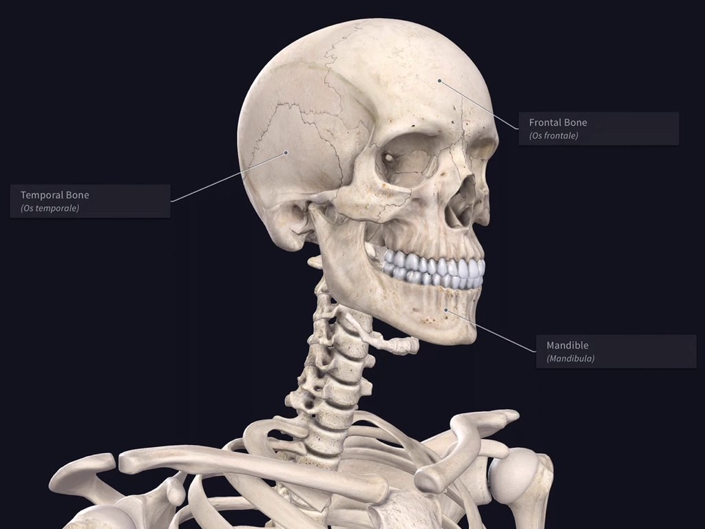
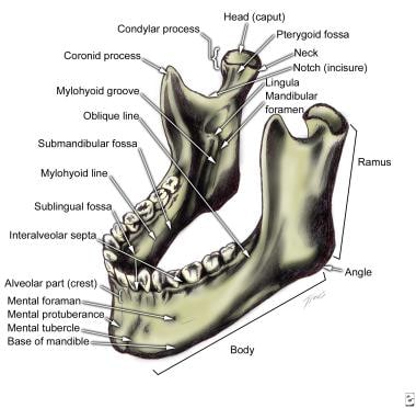
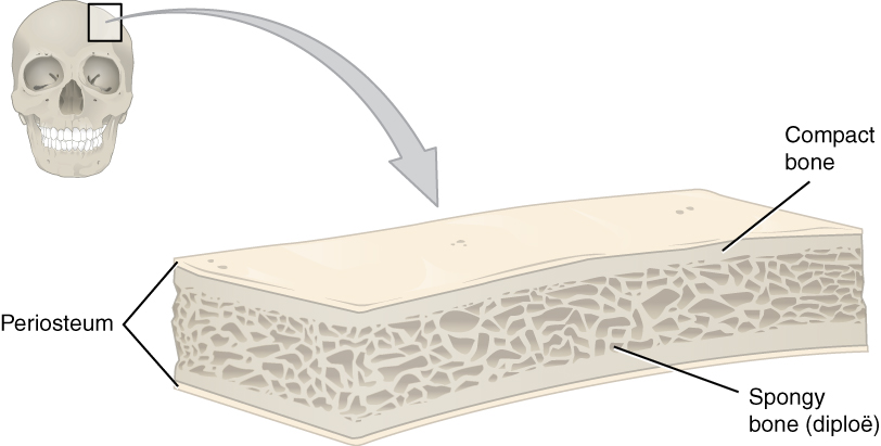
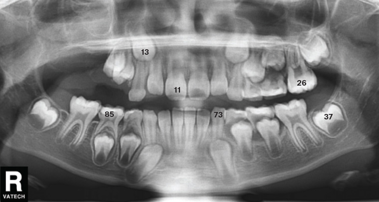
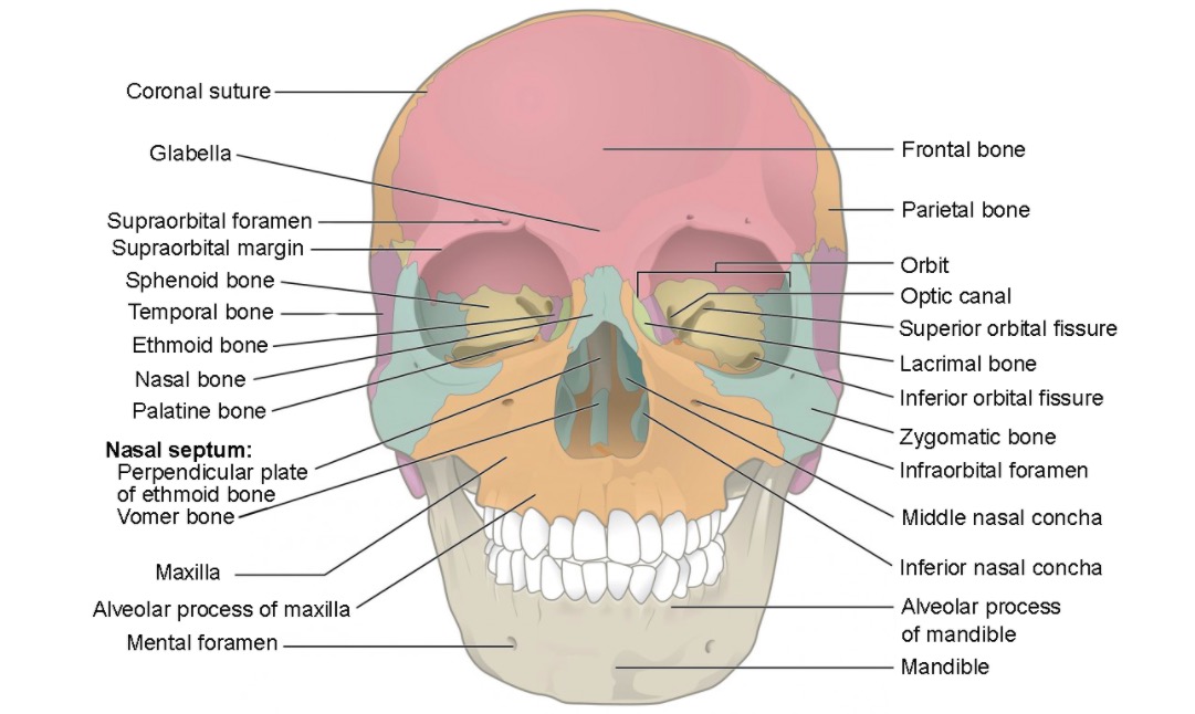
45 correctly label the following anatomical parts of the mandible.
(Get Answer) - Correctly label the following anatomical parts of ... An automobile chassis is the steel load-bearing framework of a vehicle on which the body is mounted and consists of two main parts: the frame rails and the cross members. Three percent of the letters placed in a certain postal drop box have incorrect postage. Suppose 200 letters are mailed. Radiographic positioning terminology | Radiology Reference Article ... Body positions. erect: either standing or sitting. decubitus : lying down. supine : lying on back. Trendelenburg position: the patient is supine (on an inclined radiographic table) with the head lower than the feet. prone : lying face-down. lateral decubitus : lying on one side. right lateral: right side touches the cassette.
Bones of the Foot - Tarsals - Metatarsals - TeachMeAnatomy The talus is the most superior of the tarsal bones. It transmits the weight of the entire body to the foot. It has three articulations: Superiorly - ankle joint - between the talus and the bones of the leg (the tibia and fibula). Inferiorly - subtalar joint - between the talus and calcaneus.
Correctly label the following anatomical parts of the mandible.
Temporomandibular joint (TMJ): Anatomy and function | Kenhub The temporal bone forms the superior part of the joint with two components: mandibular fossa and articular tubercle. The inferior part of the joint is formed mainly by the head of the mandible. The head of the mandible, also known as the mandibular condyle, is the posterior end of the ramus of the mandible. Head and neck: Regions and anatomy | Kenhub The submandibular triangle is a glandular area between the inferior border of the mandible and the anterior and posterior bellies of the digastric muscle. The floor of his triangle is formed by the mylohyoid and hypoglossus muscles and the middle constrictor muscle of the pharynx. The submandibular gland nearly fills this triangle. Carotid triangle (Get Answer) - Transcribed image text: 63) Creatine ... - Transtutors A) capitate B) lunate C) scaphoid D) trapezoid E) trapezium 67) The humerus A) is the most distal bone of the upper extremity B) is the longest and largest bone of the upper extremity C) articulates with the lateral end of the clavide D) is most often fractured at the anatomical neck E) may be recognized by the distinct, large trochanters at ...
Correctly label the following anatomical parts of the mandible.. Larynx: Anatomy, Function, and Treatment - Verywell Health The larynx sits at the front of the neck between the third and seventh neck vertebrae (C3 to C7), where it's suspended in position. 2 The upper portion of this organ is attached to the lower portion of the pharynx, or throat, via the hyoid bone. Its lower border connects to the upper portion of the trachea (also known as the windpipe ... 1.2.2 Skeleton Scavenger Hunt - StudyMode Write the identifying number on the Maniken® in pencil and then write the corresponding number and label on your graphic organizer. Those marked with an asterisk (*) should already be labeled. Skull (*) Mandible (*) Sternum Radius Phalanges Rib Cage Tibia Fibula Vertebral Column (cervical, thoracic, lumbar, sacral and coccyx) Scapula Carpals The mandible: Anatomy, structure, function | Kenhub The mandible is a horseshoe shaped bone of the viscerocranium. It consists of the body and two rami, connected at the angle of mandible. Body The body of mandible is its horizontal portion. It consists of two parts: The alveolar part The base of mandible The alveolar part is the upper portion of the body. What are the 12 cranial nerves? Functions and diagram - Medical News Today The cranial nerves are a set of twelve nerves that originate in the brain. Each has a different function responsible for sense or movement. The functions of the cranial nerves are sensory, motor ...
The Muscles of Facial Expression - Orbital Group - TeachMeAnatomy Anatomically, they can be divided into upper and lower groups: The lower group contains the depressor anguli oris, depressor labii inferioris and the mentalis. The upper group contains the risorius, zygomaticus major, zygomaticus minor, levator labii superioris, levator labii superioris alaeque nasi and levator anguli oris. Insect Mouth Parts & Types | Diagram, Structure & Function - Video ... The labrum is used to hold the food as it is being eaten. Mandibles: Unlike humans, which are characterized by a single lower jaw, the mouth of an insect consists of a pair of jaws, known as the... These Are the 12 Cranial Nerves and Their Functions - Healthline This division communicates sensory information from the middle part of your face, including your cheeks, upper lip, and nasal cavity. Mandibular. The mandibular division has both a sensory and a... The Pelvic Girdle - Structure - Function - TeachMeAnatomy Posterior - sacral promontory (the superior portion of the sacrum) and sacral wings (ala). Lateral - arcuate line on the inner surface of the ilium, and the pectineal line on the superior pubic ramus. Anterior - pubic symphysis.
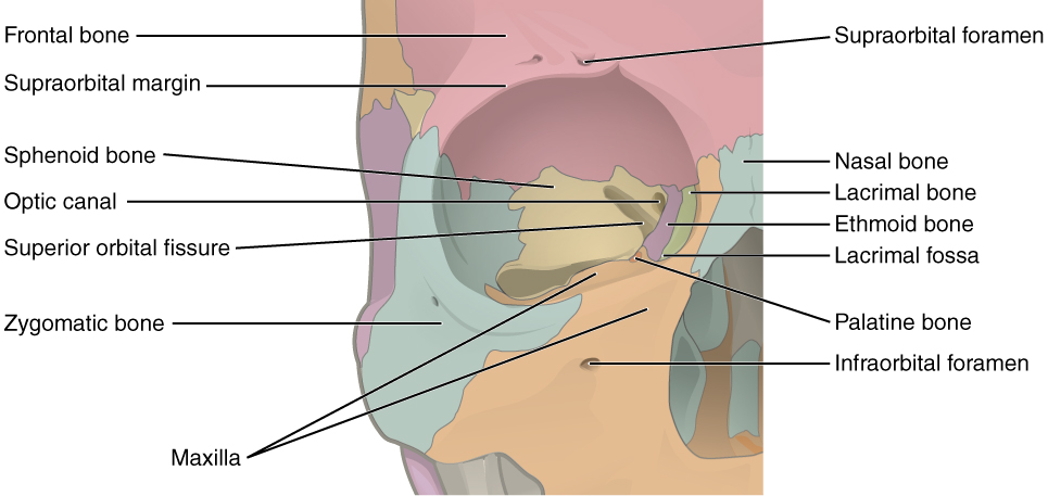
Label the bones of the orbit shown in this cadaver image by clicking ... Label the bones of the orbit shown in this cadaver image by clicking and dragging the labels to the correct location. (Some labels will not be used.) Lacrimal bone Occipital bone Sphenoid bone Maxilla Parietal bone Frontal bone Ethimold bone Mandible Zygomatic bone CEP 2016 Pedrer. Anatomy And Physiology Lab Quiz 1 - ProProfs Quiz A. Prone B. Fetal Position C. Anatomical Position D. Supine 2. What is the name of the highlighted region? A. Neck B. Anterior Cervical Triangle C. Cervical Region D. Vertebral Region 3. What makes up the lateral border of this region? A. Chin B. Mandible C. Sternocleidomastoid Muscles D. Deltoid Muscle 4. Which direction is indicated in the image? Tooth anatomy: Structure, parts, types and functions | Kenhub The tooth is made up of a crown and either single or multiple roots. The anterior teeth in both the upper and the lower jaws, from the right first premolar to the left first premolar, are single rooted teeth. On the upper jaw, the maxillary second premolar may have two roots and all of the maxillary molars have two to three roots. The Mandible: Anatomy, Function, and Treatment - Verywell Health The mandible is the large bone that holds the lower teeth in place. Also known as the lower jawbone, the mandible is the largest and strongest bone of the face. Tasked with holding the lower set of teeth in place, this bone has a symmetrical, horseshoe shape. Not directly connected to other bones of the skull, the mandible is the only moving ...
23 Blood Vessels Quizzes Online, Trivia, Questions & Answers - ProProfs A. Narrowing or complete occlusion of the vessel lumen B. Weakness of the blood vessel wall C. Damage to the adventitia D. Damage to the small arteries that prevents proper vasoconstriction and vasodilation. A and B. A, B and C.
Mastering A&P #7 - Flashcards | StudyHippo.com a. an oblique muscle layer b. a lining of columnar epithelium c. mucus-forming cells d. a circular muscle layer. answer. a All areas of the alimentary canal have a circular and a longitudinal layer of muscle. The stomach has an additional oblique layer of muscle for "wringing" itself while processing food.
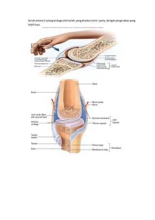
skeletal system quiz anatomy and physiology 1. Skeletal system, axial skeleton, appendicular skeleton, hip, shoulder, knee, and elbow joint Learn with flashcards, games, and more — for free. 1. 3 - the cell: learn the anatomy of a typical human cell. C) dorsal and ventral. Free Anatomy Quiz Anatomy Of The Hand Bones Quiz 1. hpassons85_74484.
Anatomy, Head and Neck, Mandible - StatPearls - NCBI Bookshelf The mandible is the largest bone in the human skull. It holds the lower teeth in place, it assists in mastication and forms the lower jawline. The mandible is composed of the body and the ramus and is located inferior to the maxilla. The body is a horizontally curved portion that creates the lower jawline. The rami are two vertical processes located on either side of the body; they join the ...
Oral cavity: Anatomy, tongue muscles, nerves and vessels | Kenhub The tongue is the central part of the oral cavity. It's a muscular organ whose base is attached to the floor of the oral cavity, whilst its apex is free and mobile. The tongue is predominantly muscle. There are 8 in total; 4 intrinsic muscles and 4 extrinsic. For more on these muscles, see the tongue muscles section below.
Anatomy, Bone Markings - StatPearls - NCBI Bookshelf Tibial and femoral condyles are palpated to approximate the menisci sites during the McMurray test, which evaluates the structural integrity of the meniscus. Bony landmarks of the elbow are used to orient the operator and locate areas of interest for targeted medical imaging like ultrasound.
Muscles of the Vertebral Column: Support & Movement The erector spinae is a group of muscles that work together to extend the vertebral column and thus maintain good posture. The muscles are innervated by the spinal nerves. Many of these muscles ...
Correctly Label The Following Anatomical Parts Of A Flat Bone The anatomical parts of a flat bone are named according to their location within the body. The superior portion of the bones is above another part of the body and the inferior part is below it. This is the same way for the limbs and muscles in humans. The upper arm is called the antebrachium, and the lower arm is called the femur.
Mandible - ProProfs Quiz The femur is the strongest bone in the human body. It extends from the hip to the knee. It has the ability to resist a force of up to 1,800 to 2,500 pounds before it can break. Take up the quiz below and see how much you know...
The Arches of the Foot - Longitudinal - Transverse - TeachMeAnatomy Fig 1 - The transverse, medial longitudinal and lateral longitudinal arches of the foot. Longitudinal Arches There are two longitudinal arches in the foot - the medial and lateral arches. They are formed between the tarsal bones and the metatarsal heads Medial Arch The medial arch is the higher of the two longitudinal arches.
Penis: Anatomy, Function, Disorders, and Diagnosis - Verywell Health It consists of several parts, including the shaft, head, and foreskin.
(Get Answer) - Transcribed image text: 63) Creatine ... - Transtutors A) capitate B) lunate C) scaphoid D) trapezoid E) trapezium 67) The humerus A) is the most distal bone of the upper extremity B) is the longest and largest bone of the upper extremity C) articulates with the lateral end of the clavide D) is most often fractured at the anatomical neck E) may be recognized by the distinct, large trochanters at ...
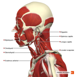
Head and neck: Regions and anatomy | Kenhub The submandibular triangle is a glandular area between the inferior border of the mandible and the anterior and posterior bellies of the digastric muscle. The floor of his triangle is formed by the mylohyoid and hypoglossus muscles and the middle constrictor muscle of the pharynx. The submandibular gland nearly fills this triangle. Carotid triangle
Temporomandibular joint (TMJ): Anatomy and function | Kenhub The temporal bone forms the superior part of the joint with two components: mandibular fossa and articular tubercle. The inferior part of the joint is formed mainly by the head of the mandible. The head of the mandible, also known as the mandibular condyle, is the posterior end of the ramus of the mandible.















Post a Comment for "45 correctly label the following anatomical parts of the mandible."