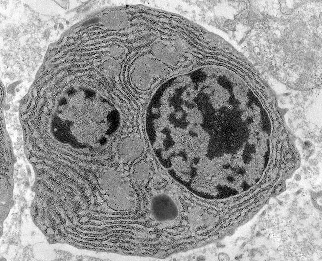
38 transmission electron micrograph labeled
Solved Label this transmission electron micrograph of | Chegg.com Anatomy and Physiology questions and answers Label this transmission electron micrograph of relaxed sarcomeres by clicking and dragging the labels to the correct location Sarcamere 1 band (light) Z disc Mline Aband (dark) H zone Transmission Electron Microscope (TEM) - Uses, Advantages and Disadvantages A Transmission Electron Microscope produces images via the interaction of electrons with a sample. TEMs are costly, large, cumbersome instruments that require special housing and maintenance. They are also the most powerful microscopic tool available to-date, capable of producing high-resolution, detailed images 1 nanometer in size. ...
Transmission Electron Microscope (With Diagram) The final image in a TEM is known as transmission electron micrograph. The salts of some heavy metals, e.g., lead; osmium, tungsten and uranium are often used for staining. These heavy metal stains are used to increase the contrast between ultra structures and the background.

Transmission electron micrograph labeled
Transmission electron microscopy DNA sequencing - Wikipedia Transmission electron microscopy (TEM) produces high magnification, high resolution images by passing a beam of electrons through a very thin sample. Whereas atomic resolution has been demonstrated with conventional TEM, further improvement in spatial resolution requires correcting the spherical and chromatic aberrations of the microscope lenses. Transmission Electron Microscopy | MRSEC The Electron Microscopy Facility is a joint BSD/PSD resource available to all campus researchers. Users have access to an FEI Tecnai F30 scanning/transmission electron microscope. The microscope has a point-to-point resolution of 0.2 nm when operated in the TEM mode and a spatial resolution of 0.2 nm for the STEM mode.The facility is located in ... Microscope Types (with labeled diagrams) and Functions The shorter wavelength of electrons compared to visible light photons helps the observer achieve a very high resolving power compared to normal microscopes thereby aiding observers to see very tiny objects clearly. Electron microscope labeled diagram The different types of electron microscopes are: Transmission Electron Microscope
Transmission electron micrograph labeled. Electron Microscope- Definition, Principle, Types, Uses, Labeled Diagram There are two types of electron microscopes, with different operating styles: 1. Transmission Electron Microscope (TEM) The transmission electron microscope is used to view thin specimens through which electrons can pass generating a projection image. The TEM is analogous in many ways to the conventional (compound) light microscope. Transmission Electron Microscope (TEM)- Definition, Principle, Images May 19, 2022 · The working principle of the Transmission Electron Microscope (TEM) is similar to the light microscope. The major difference is that light microscopes use light rays to focus and produce an image while the TEM uses a beam of electrons to focus on the specimen, to produce an image. PDF Identifying Organelles from an Electron Micrograph The photograph shown below details chloroplast structure as viewed with a transmission electron microscope Courtesy of Dr. Julian Thorpe - EM & FACS Lab, Biological Sciences University Of Sussex A single Granum Chloroplast envelope visible as two membranes Stroma containing numerous small ribosomes Lamellae connecting different grana Looking at the Structure of Cells in the Microscope Determining the detailed structure of the membranes and organelles in cells requires the higher resolution attainable in a transmission electron microscope. Specific macromolecules can be localized with colloidal gold linked to antibodies. Three-dimensional views of the surfaces of cells and tissues are obtained by scanning electron microscopy.
Transmission Electron Microscopy - Penn State College of Medicine Research The JEOL 1400 TEM (Room C1727) is capable of generating ultra-structural nanoscale images from fixed cell/tissue samples or multiplexed immune-labeled samples. Computer-controlled operations Resolution up to 3 Angstroms Magnification up to 370,000X Capable of collecting data suitable for 3D reconstructions of negative-stained samples Transmission Electron Microscope: Definition, Parts, Working Principle ... In a Transmission electron microscope, the electron beam is transmitted through a very thin specimen or object and forms a highly magnified and detailed image of the sample. This microscope uses electron beams instead of light. The specimen used in Transmission Electron Microscope, should be very thin, less than 100 nm thick. Label This Transmission Electron Micrograph Of A Relaxed Sarcomere ... Label this transmission electron micrograph of relaxed sarcomeres by clicking and dragging the labels to the correct location . Label the following image using the terms provided. Note how the sarcomeres are extended to only approximately 120 % . IMG_2132 - FIGURES Label this transmission electron from anatomy 10.png - Label the transmission electron micrograph... anatomy 10.png - Label the transmission electron micrograph of the. anatomy 10.png - Label the transmission electron micrograph... School Utah Valley University; Course Title ZOOL 1090; Uploaded By emileeroylance19. Pages 1 Ratings 67% (3) 2 out of 3 people found this document helpful;
Transmission Electron Microscopy - an overview | ScienceDirect Topics platelet transmission electron microscopy (tem) is an essential tool for laboratory diagnosis of various hereditary platelet disorders since it was first used to visualize fibrin-platelet clot formation in 1950s.51,52 platelet tem employs two main methods to visualize platelet ultrastructure, whole-mount tem and thin-section tem. 53,54 … Transmission Electron Microscopy (TEM) - Warwick The transmission electron microscope is a very powerful tool for material science. A high energy beam of electrons is shone through a very thin sample, and the interactions between the electrons and the atoms can be used to observe features such as the crystal structure and features in the structure like dislocations and grain boundaries. Transmission electron microscopy techniques, Electron microscopy, Otago ... The small black dots label the dendritic arms of two GnRH neurons (pseudocolored green and purple), cells in the brain that control fertility. ... STEM (scanning transmission electron microscopy) enables the use of other of signals that cannot be spatially correlated in TEM, including secondary electrons, scattered beam electrons ... Transmission electron microscopy (TEM) The mPrep System™ streamlines Transmission Electron Microscopy (TEM) sample preparation using a capsule based approach. Use mPrep/s capsules to fix, orient, embed, and section specimens. Use mPrep/g capsules to stain or immuno-label TEM grids.. The mPrep System efficiently produces quality results from every sample.
Transmission electron microscopy - Wikipedia Transmission electron microscopy (TEM) is a microscopy technique in which a beam of electrons is transmitted through a specimen to form an image. The specimen is most often an ultrathin section less than 100 nm thick or a suspension on a grid. An image is formed from the interaction of the electrons with the sample as the beam is transmitted through the specimen.

Transmission electron micrograph of animal cell - Stock Image - G450/0051 - Science Photo Library
Labeling the Cell Flashcards | Quizlet Label the transmission electron micrograph of the nucleus. membrane bound organelles golgi apparatus, mitochondrion, lysosome, peroxisome, rough endoplasmic reticulum nonmembrane bound organelles ribosomes, centrosome, proteasomes cytoskeleton includes microfilaments, intermediate filaments, microtubules Identify the highlighted structures
Electron Micrographs** Below is a collection of electron micrographs with labelled subcellular structures that you should be able to identify. Also, be sure to observe any electron micrographs which are made available in the laboratory by the instructor.
Transmission electron microscopy DNA sequencing - Google Search Transmission electron microscopy DNA sequencing is a single-molecule sequencing technology that uses transmission electron microscopy techniques.The method was conceived and developed in the 1960s and 70s, but lost favor when the extent of damage to the sample was recognized. DNA is visible under the electron microscope; however, it must be labeled with heavy atoms so that the DNA bases can be ...
Label This Transmission Electron Micrograph : TEM of chloroplast from ... Label this transmission electron micrograph of relaxed sarcomeres by clicking and dragging the labels to the correct location . Transmission electron microscopy (tem) is one of the oldest technologies and still. Molecular labeling for correlative microscopy: Fluorescence microscopy in combination with tem and an ion beam analysis (iba, which ...
The Transmission Electron Microscope | CCBER Transmission electron microscopes (TEM) are microscopes that use a particle beam of electrons to visualize specimens and generate a highly-magnified image. TEMs can magnify objects up to 2 million times. In order to get a better idea of just how small that is, think of how small a cell is.
Transmission Electron Microscopy: Theory & Applications A transmission electron microscope (TEM) is a special type of microscope that uses electrons for magnification. The magnification in a standard optical microscope is limited by the wavelength of ...
Transmission electron microscopy characterization of fluorescently ... Transmission electron microscopy characterization of fluorescently labelled amyloid β 1-40 and α-synuclein aggregates Abstract Background: Fluorescent tags, including small organic molecules and fluorescent proteins, enable the localization of protein molecules in biomedical research experiments.


Post a Comment for "38 transmission electron micrograph labeled"